FIGURE 42-A: The incision on the cerebral hemispheres that are to be transplanted is begun.
(this material is an excerpt from the book "The Message of the Stones", by Dr. Javier Cabrera)
(EXCERPT #5B)
TRANSPLANT OF THE CEREBRAL HEMISPHERES
Contemporary medicine has not yet been able to transplant a human brain. The experiments in this field have been limited to animals. It has been possible to isolate a monkey brain and maintain it alive for a relatively long period of time. There have also been attempts to transplant monkey's heads, but with discouraging results. Until recently, the major problem was to figure out how the nervous fiber, after being cut, could regenerate itself and recuperate its regular function. It is true that there have been cases where human nerves have been sutured, especially in accident injuries, but the sutures have been made to join nerves from the same organism, and even in this area the techniques employed have not been effective in other cases. A recent experience offers hope for the future: in rats given, special treatment before and after the rupture of the spinal chord, it has been proved - after the union of the segments of the spinal cord from the same animal - the regeneration of the nerve fiber (axon) and the recovery of the nerve impulse. The experiment was done by five investigators from the University of Michigan, U.S.A. Earl R. Feringa, Gary G. Gurden, William Strodel, William Chandler and James Knake. The rats used in these experiments were female albinos crossed among brothers for mare than sixty-two generations, and were immunized through the application of an antigen of the spinal medulla from rats of the same blood. Access to the writings which contain information about this experiment (35) was made available to me through the kindness of Dr. Jorge Voto Bernales, eminent Peruvian neurologist, under whom I had the honor of studying at the Universidad Nacional Mayor de San Marcos and the pleasure of working with in the Seguro Social of Peru.
The importance of the experiment described above seems to me to be in that such regeneration has been achieved in the spinal medulla - something which has never before been achieved - due to the fact that the spinal medulla is composed of numerous nervous fibers. In addition, the importance of the experiment lies in the treatment given the rats, for this allows us to suppose that we are on the way to developing a technique that would permit a control of the factors that determine the possibility of cutting and regenerating nervous fibers. There is something that strikes me as particular about this experiment: the use of female rats. The researchers do not indicate their motives for having made this choice of specimens. What has been already mentioned about the knowledge that contemporary medicine already has about the suprarenal gland growing in size at the beginning of the menstrual cycle and during pregnancy should be remembered. Also recall that I have attributed this phenomenon to the increase in the excretion of the hormone that stimulates the cells of the organism so that it may tolerate the reception of a foreign element, the spermatozoid, and then the fetus. I believe that the success of this experiment must be due to the fact that the female rats, due to their great fecundity and short period of gestation, must have a high level of this hormone in their blood, for, if due to this hormone the female can tolerate foreign tissue, then more so will she be able to regenerate her own cells, in this case the nervous fibers that are nothing but the extension of a part (citoplasma) of the nerve cells.
Despite the great advances of contemporary neurosurgery, the day when a complete brain can be transplanted is far off. For this there must exist infallible techniques to avoid rejection of the transplant and to assure the regeneration of the nervous fiber, and it is also necessary to have a complex system of apparatus, probably electronic, not only to facilitate the transplant operation, but also so that this system nay assure control over all the biological functions of the individual whose brain must be extracted so that another may be implanted. This complex system will temporarily replace the human brain that is to be replaced and the human brain that will replace it gliptolithic surgery and such a complex system of apparatus and they used it in all the surgical operations that are seen on the gliptoliths. With this apparatus it was possible to avoid all the risks that the patient to be operated on was subject to, such as a cessation of the breathing functions, a cessation of the heart, different types of shocks, etc. We are dealing with a system represented through symbols inscribed on the instruments, on the operating table, on the body of the patient and on the surgeons and organs. Gliptolithic surgery informs us about this complex system of apparatus only to the extent that it refers to the type of instruments used and to the general functions they perform, and not their physical nature.
Before going on to describe the technique used by the gliptolithic surgeons to transplant cerebral hemispheres, I think it opportune to go over some of the basic aspects of the anatomy of the nervous system. It consists of two well distinguished parts: cerebrum or brain - enclosed in the bony box known as cranium - and the spinal chord enclosed in the vertebral canal or foramen of the backbone. The cerebrum or abrain is composed of several parts; two cerebral hemispheres (left and right), the thalamus, cerebellum, the pons, cerebral peduncles and the racheos bulb. The cerebrum has two lateral egg-shaped and symmetrical masses called hemispheres. They are covered by the cerebral cortex composed of gray matter. It is in the cerebral cortex where the sensory organs are converted into conscious sensations, and it is also here where the centers that initiate and control voluntary movements are found. All this makes it possible to affirm that it is in the cerebral cortex where the functions take place that allow an individual to acquire and retain knowledge. It is constituted by cells that are particular in that they do not regenerate. The deeper layers of the cerebral hemispheres, also called the white matter, is constituted by nerve fibers (extensions of the nervous cells), through which the nervous impulses come and go to the cerebral cortex, and from the cortex of one hemisphere to the cortex of another. The two symmetrical ovals that make up the cerebral hemispheres are united in the middle by a strong bridge of nervous fibers (white substance). This bridge is called the corpus callosum and under it the thalamus is located. This section, and the others that constitute the rest of the brain - the cerebellum, protruberance, peduncles and the bulb - contain the centers that command the functioning of the organs of an individual, that is, the vegetative life. Under the bulb - at the lowermost extreme of the brain - and at the level of the occipital orifice, begins the spinal chord. From all the elements I have just described the cranial pairs grow, consisting of twelve pairs of nerves that are distributed principally through various organs and tissues in the head and the neck.
To transplant this complex of the nervous system that is lodged in the cranial cavity, that is the brain, is the object of the research that has been made recently in the area of experimental neurosurgery. This presupposes the necessity of cutting the spinal medulla close the racheous bulb and then achieving the regeneration of the nerve fiber at the place of the cut after the transplant has been finished. But it also presupposes - and this makes the operation extremely difficult - cutting and regenerating the fibers of the twelve pairs of cranial nerves. It is possible that these complications nay have led to an attempt to transplant a whole head instead of just a brain, a method that has been named cephalotransplant and that has been attempted in monkeys, with discouraging results as I have already mentioned. I think that a more advance stage - one that contemporary neurosurgery has not yet attempted - is the transplant of the cerebral hemispheres, that implies cutting the bridge that unites then, the corpus callosum, which being made up of nervous fibers may be able to regenerate itself (remember the experiment with the rats that demonstrates that nervous fiber can regenerate itself). It also implies the cutting and suturing of the main arterial and veinous blood lines that irrigate the cerebral hemispheres with blood. The cutting of the corpus callosun should not interfere with the thalamus, for here are very important nervous functions. Contemporary surgery has one type of operation in which one of the cerebral hemispheres is removed. To do this an incision is made at the level of the corpus callosum without affecting the thalamus. This operation is made in order to improve the mental capacity of retarded children, to reestablish movement for people who have, experienced partial paralysis due to cerebral hemorrhages, etc. In many cases it has been successful.
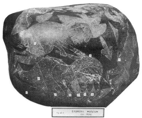
The brain transplant referred to in the gliptoliths in my possession is of the cerebral hemispheres. The fact that the cerebral cortex is part of the cerebral hemispheres and that in it are located the cognitive functions of the brain, and knowing that the gliptolithic humanity has as a goal of its existence the development of the reflexive capacity to increase and conserve knowledge, it is not surprising that they attempted to transplant the hemispheres of the brain. The information as to this type of transplant is contained in a series of eleven gliptoliths. Transplanted into another person's head. The operating table, represented symbolically, (1 in Fig. 42A), has parallel and oblique lines inscribed upon it whose direction will change in future scenes solely for the purpose of indicating that the scene corresponds to another individual. On top of the table appear, connected to the individual in different parts of the body, some segments with rhomb-shaped sections, symbols of animal life, which in this case suggests those biological functions that occur in the human organism without the control of the individual (vegetable functions). These segments with rhomb-shaped figures represent symbolically elements of that complex system of electronic apparati - to with I have previously referred - that are serving in this case to stimulate and maintain uninterrupted these vegetative functions: stimulus and control of respiration, the diffusion of nutritive elements, stimulus and control of the cardiovascular system, stimulus and control of the digestive and glandular system (5, 6, 7 and 8, respectively, in Fig. 42A). The triangular figure full of rhomb shapes at the foot of the patient is a symbol of biological animal energy and therefore of unconsciousness, that indicates here that the patient cannot feel anything and is perfectly still. The signs engraved on the clothes of the patient and the surgeon indicate the intensity of the electronic energy that this complex system of apparati needs to stimulate and maintain the vegetative functions during the operation. Thus, it the signs are squares, this indicates the highest intensity of energy; if they are rhomb-shaped this signifies a little less intensity; if they are parallel lines, this signifies a much lower level of energy (3 and 15 in Fig. 42A). If the patient is not wearing clothes (pants), this indicates that the minimum of energy needed to keep him alive is being used.
FIGURE 42-A: The incision on the
cerebral hemispheres that are to be transplanted is begun.
In another scene on the same gliptolith the moment at which the surgeon extracts the cerebral hemispheres from the same individual can be seen (9 in Fig. 42B). Observe the direction of the parallel lines on the operating table; it is the same as the previous scene, indicating that we are dealing with the same individual, but now in a state of deep relaxation judging by the looseness of his upper limbs.
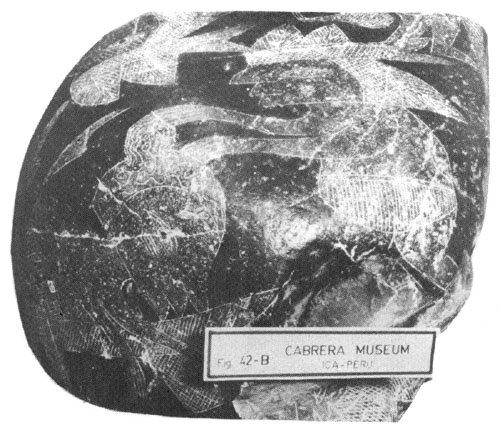
FIGURE 42-B: Extraction of the
cerebral hemispheres to be transplanted.
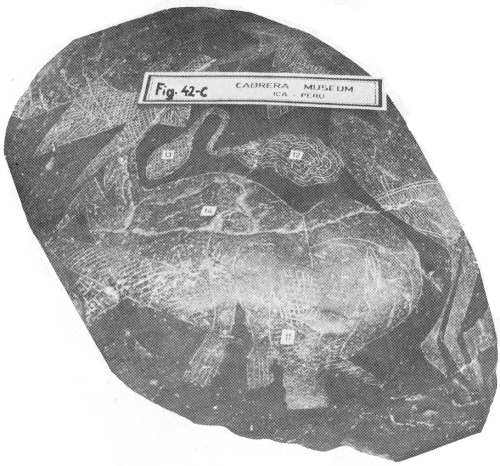
The third and last scene on this gliptolith shows the cerebral hemispheres which have already been extracted (2 in Fig. 42C), which are being irrigated, probably with blood from a pregnant woman, in order to stimulate and keep the nervous cells alive and to stimulate the regeneration of the fibers. The apparatus used for irrigation is connected to the arterial and veinous vessels that lead to the hemispheres. The rhomb-like figures that fill the base of the irrigating apparatus signify that it is being run by electronic energy of medium intensity. This stage of the operation is of vital importance, because the nervous cells cannot maintain themselves in a normal state for more than three minutes without oxygen and glucose, fundamental elements for their metabolism that are provided through the circulatory system. In this phase of the operation, the complex system of electronic apparati has been directed towards stimulating and controlling preferably the cardiovascular system of the individual, as can be deduced from the segment full of rhomb-like figures that can be seen under his neck (11 in Fig. 42C). This procedure is due to the fact that the patient is in a critical situation having had his cerebral hemispheres extracted. The absence of clothing (14 in Fig. 42C) reiterates that this is a critical situation since it reveals that a minimum of electronic energy is being used to keep him alive. Observe the direction of the parallel lines inscribed on the operating table: it expresses that the patient is the same person as in the two previous scenes.
The following gliptolith has only one scene that represents the moment when the surgeon finishes suturing the wound of the individual from whom the cerebral hemispheres have been extracted to be transplanted (2 in Fig. 43). The rhomb-like figures that fill the base of the apparatus that is used to suture indicate that this apparatus is run by medium intensity electric energy. The segments full of squares that can be observed under the face and the neck of the patient (5 in Fig. 43), signify that the maximum level of electronic energy is being used to activate and maintain all the biological functions of the individual, since he has been deprived of the essential centers of cerebral activity, and thus of consciousness (as can also be seen by the symbol that appears at the foot of the patient) (6 in Fig. 43) The direction of the lines on the operating table indicate that this is the same patient as in the previous scenes. The leaf - a symbol of life - that appears under the operating table (1 in Fig. 43), signifies that the individual is living by artificial means.
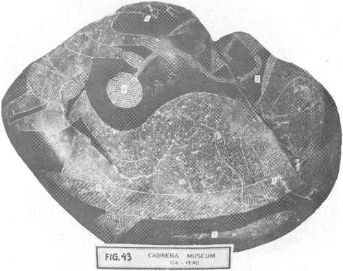
FIGURE 43: The suture of the
wound of the individual whose cerebral hemispheres have been extracted to be
transplanted.
The rest of the gliptoliths of this series show scenes of the more advanced phases of the same transplant and refer to the individual who is going to receive the cerebral hemispheres. One of these gliptoliths shows the instant in which the cerebral hemispheres are extracted from this individual (1 in Fig. 44). The change in direction of the lines on the operating table indicates that this is a different individual from the one appearing in the previous scenes. In this phase of the operation the danger of heart failure is symbolized by the segment of rhomb-like figures under the neck (4 in Fig. 44) that signifies that the complex system of electronic apparati has concentrated its actions on the stimulation and control of the individual's cardiovascular system. The rhomb-like figures on the clothes (pants) of the individual (5 in Fig. 44) indicate the use of a medium intensity electronic energy in this phase. The triangular segment full of rhomb-like figures (2 in Fig. 44) that can be observed under the eye, signifies that the individual is ignorant of the operation not because he is anaesthetized but rather because his cerebral hemispheres have been removed.
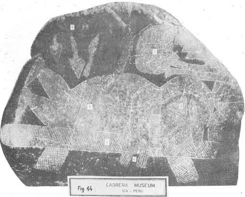
FIGURE 44: The instant at which
the cerebral hemispheres of the individual are extracted to be replaced by
others.
Another gliptolith shows the next scene: the continuation of the irrigation of the cerebral hemispheres that were extracted from the first individual and that are now to be transplanted (Fig. 45) this scene shows a surgeon irrigating the hemispheres, using an apparatus already described in a previous scene (5 in Fig. 45).
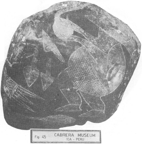
FIGURE 45: The irrigation with
blood of the cerebral hemispheres continues before they are transplanted.
The next gliptolith of the series has two scenes. In the first there is a particularly interesting feature: the direction in which the parallel lines on the operating table are pointing (2 in Fig. 46A) is the same as in the scenes that show the individual from whom the cerebral hemispheres were extracted to be transplanted into another. This could lead one to believe that the individual in this scene is not going to receive the transplant. However, the direction of the parallel lines indicates that it is the cerebral hemispheres in this scene that belong to the previous individual, that have just been transplanted into the second individual (1 in Fig. 46A). With the particular use of the symbols on the table I understand that the surgeons of the gliptolithic culture are attempting to express that the man is where his cerebral hemispheres are. The symbolic presence of the first individual in this scene is reiterated by the leaf that can be seen in the lower part (4 in Fig. 46A). In this case the leaf, - symbol of human life - signifies life capable of reflection, and its particular position signifies that this reflective life has arrived in the cerebral hemispheres of the first individual to be united with the biological life of the second individual. The leaf that is engraved on the upper part of the scene (5 in Fig. 46A) signifies that the body is alive, and the segments full of rhomb-like figures under the face and the neck indicate that the biological functions of this body are being stimulated and controlled by the aforementioned complex system of electronic apparati.
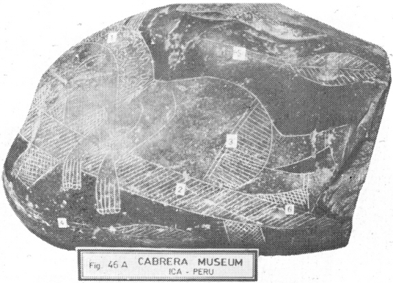
FIGURE 46-A: The brain
hemispheres were just transplanted.
The other side of the gliptolith that I have just described will confirm beyond a doubt that the transplant has been of the cerebral hemispheres. It is to be observed that this is the only gliptolith of the whole sequence that I have described that shows the irrigating apparatus with two channels, which indicates that both hemispheres, not just one, are being irrigated.
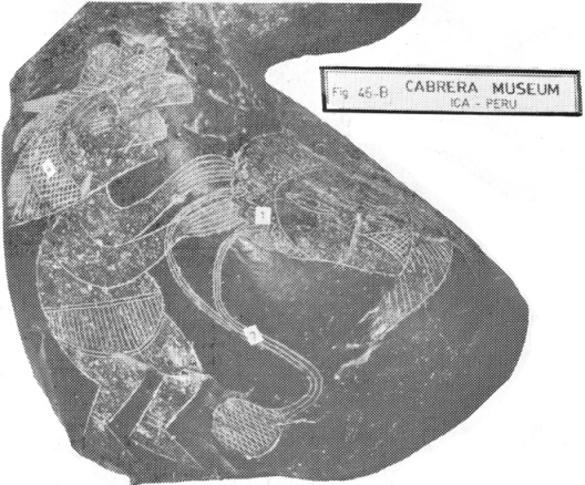
FIGURE 46-B: The two channels of
the blood irrigating mechanism reveal that not one but both cerebral hemispheres
have been transplanted.
The last gliptolith of this series indicates that the operation has come to an end. Two surgeons can be seen finishing the suture of the wound of the individual that has received the cerebral hemispheres (7 in Fig. 47). The instrument used to suture has two branches that are attached to the surgeon's foreheads. Since the surgeons have two half-leaves on their heads - a symbol of the apparati that allows the gliptolithic humanity to capture and convert solar (photonic) energy and cosmic (corpuscular) energy into electronic energy - it is understood that the suture apparatus is activated by electronic energy. The presence of the leaf symbol in the arms of one of the surgeons indicates that the branch of the suturing instrument that he is holding in his hand is the one that used electronic energy and that the other one is only a ground wire. The rhomb-like figures on the circular section of the instrument reveal that the electronic energy that is used is of medium intensity. The rhomb-like figures connected to different parts of the patient should also be pointed out. They are, once more, the symbol of the fact that the biological functions of the patient are being stimulated and controlled by the complex system of electronic apparati. The rhomb-like figures indicate that the electronic energy is of medium intensity.
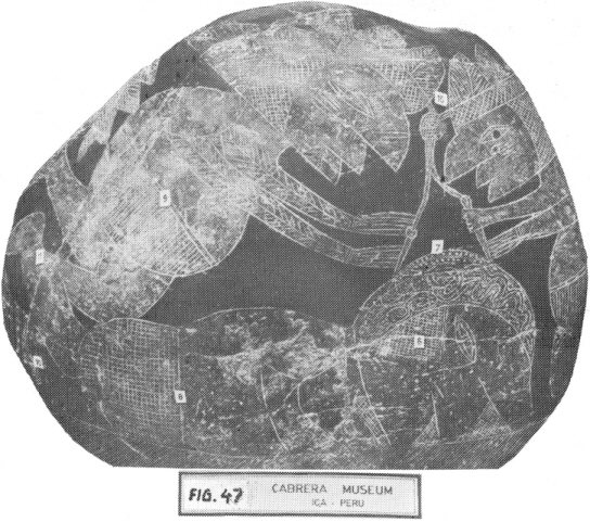
FIGURE 47: The transplant of the
cerebral hemispheres has come to an end: the surgeons
suture the wound of the individual who has received the transplanted
hemispheres.
It is possible that the electronic energy used through the operation may have come from the apparatus symbolized by the half-leaves that the surgeons wear on their heads, and that the surgeons may have projected this energy through themselves to the instruments and the apparati of the complex system that I have indicated is electronic. In this case, the surgeons, by determining the intensity of the electronic power, must have acted as regulators (relay) of the energy flow. This is confirmed by the presence of rhomb-like figures on the bands that the surgeons wear on their heads, as well as on the instruments and apparati that symbolically represent the stimulus and maintenance of the biological functions of the individual through the complex system of electronic apparati.
-----------------------
Footnotes:
(37) I use the word Megapolis for the big gatherings of reflexive and scientific human beings. It is of my understanding that these men used to live in the Megapolis with different kinds of men dedicated to the various activities related to the life in the planet.
(38) The alticamellus was a mammiferous that lived, according to Paleontology, 13 billion years ago in the Tertiary period. It is the equivalent to the present camel from the desert. They were adapted to high temperatures with restricted hydrological resources. The alticamellus on the stone shows us – graphically - the thermocritical situation shown by the planet. The average temperature had been increased up to such a point that zoological life was only possible for conditioned species.
(39) Confront "El origen de los primeros seres humanos". In Cuentos, mitos y leyendas del antiguo Peru. Compiled by Jose Maria Arguedas and Francisco Izquierdo Rios. Ministerio de Educacion (Board of Education) Lima, 1947.